service - Negative staining
B A C KGet a new perspective on sample quality with visual feedback
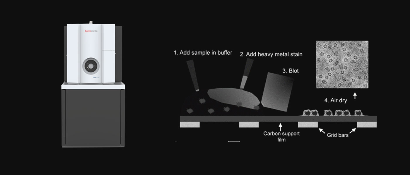
Negative staining is a convenient method to assess the quality of biological samples under the microscope for the observation of particulate matter or biological macromolecules in the sample.
After purification, the sample needs to be evaluated to see whether it is sufficiently pure and homogeneous for further, more detailed study by electron microscopy.Qualified samples should show homogeneous size, no aggregation, separated as expected.

Negative staining Workflow
Why do you need Negative staining Services?
● Detection of 2D characterization of biomolecules such as proteins
● Detection of sample purity and homogeneity
● Exploring the sample preparation conditions suitable for freezing samples
Why do you need Negative staining Services?
ShuimuBiosciences is the largest commercial cryo-electron microscopy platform in the Asia-Pacific region with a 300kV top-of-the-line cryo-electron microscope as its core. The platform is equipped with G3 Titan Krios, Talos L120C, Vitrobot and plasma processor to provide leading-edge SPA structure resolution services.
● Detection of 2D characterization of biomolecules such as proteins
● Detection of sample purity and homogeneity
● Exploring the sample preparation conditions suitable for freezing samples

About the samples
Protein (30-50ul,≥2mg/mL,Purity >95%):
SDS-PAGE without obvious stray bands and degradation bands, also to ensure that it is the target protein (mass spectrometry identification, etc.).
Buffer (50-100 ml):
SDS-PAGE without obvious stray bands and degradation bands, also to ensure that it is the target protein (mass spectrometry identification, etc.).
Homogeneity:
Before negative staining, try to do gel filtration chromatography on the sample (this step is necessary if it is a complex), if the result shows a single peak, it proves that the sample homogeneity is greater than 90%. (Glycosylation or phosphorylation modifications have less effect on the electron microscopic structure resolution); after gel filtration layer, do not concentrate again to avoid protein aggregation, etc.
Storage (10ul-20ul/tube):
It is recommended that samples be dispensed directly after preparation to avoid the need for repeated freezing and thawing during the experiment, resulting in unusable samples.