


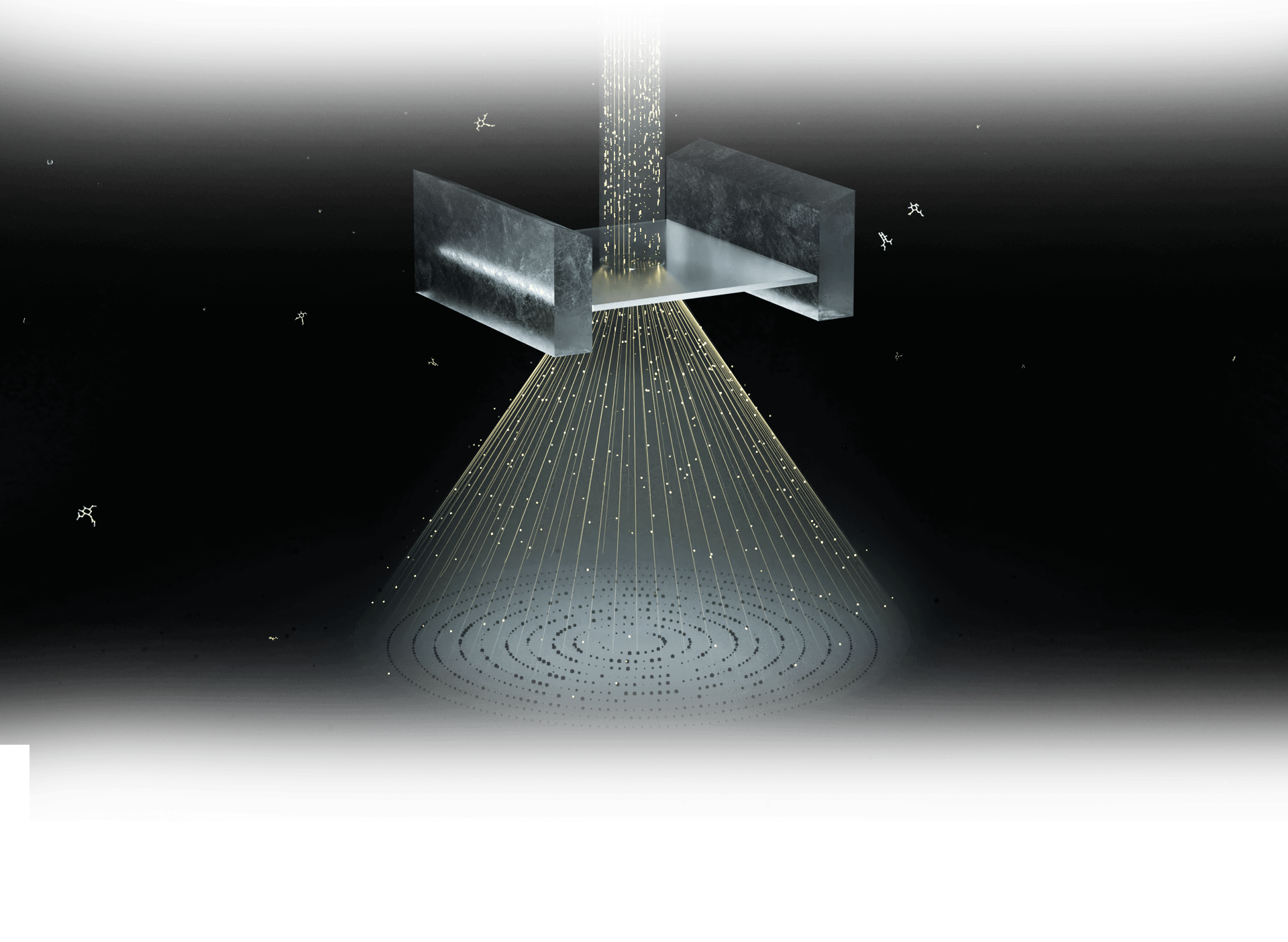
TECHNOLOGY - MicroED
B A C KeTasED Stage-camera synchronization
for conventional CryoEM
A stage-camera synchronization scheme minimizes the hardware requirements and enables the use of the conventional electron cryo-microscope with a single-frame CCD camera, which ensures not only the acquisition of ultrahigh-resolution diffraction data but also wider application of this technique
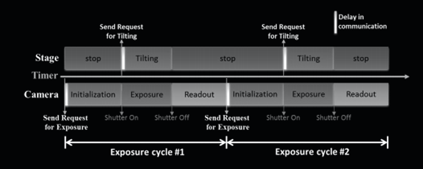
Fig1. Schematic diagram of the stage-camera synchronization.
MicroED are revealing structures at an increasing pace, though the technique is still undergoing rapid development.
Even crystallography has witnessed a dramatic reduction in the crystal size required for adequate diffraction, growth of large and high-quality crystals represents the bottleneck of structure determination by crystallography approaches. As a novel crystallographic method, MicroED exploits the advantage that electrons interact much more strongly with material and posit considerably less damaging energy into a crystal. Electron diffraction data can be collected from extremely small nanocrystals at a low dose, which benefits structure research of crystallizing problematic proteins. With this new modality in cryo-EM, novel structures considered untraceable before were successfully determined today.
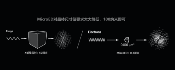
Figure 2. Crystal volume difference between MicroED and X-ray crystallography.
However, special requirement for radiation-tolerated movie-mode camera and the lack of automated data collection method increases the barrier to the utilization of MicroED. ShuimoBio drives the development of useful software to simplify and speed workflows in a way that broadens accessibility to MicroED. Pro. Xueming Li, one of our scientific advisors, has devised a stage-camera synchronization scheme to minimize the hardware requirements, which will accelerate the adoption of best practices for access.
Figure 2. Crystal volume difference between MicroED and X-ray crystallography.
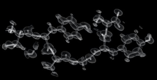
Figure 3. Structures solved from the merged datasets.
Benefiting from eTasED, microED can be realized on a conventional cryo-EM system without any modification
Published work using MicroED are still limited to several groups given to the requirement of specific camera hardware and high-end microscopes. Development of stage-camera synchronization scheme will advance the field and demystify more crystal structure.
- Tailored for single-frame camera. The stage tilting and sample illumination are activated and inactivated at predefined time points so that they are only activated within the effective exposure step of an exposure cycle.
- Available for movie-mode camera. With a movie-mode camera, the exposure time can be set much longer than that of a single-frame camera, such that each tilting-exposure cycle covers a large or even entire range of the tilting angle.
- Compatible with 120kV TEM. Ultrahigh-resolution diffraction data of peptide nanocrystals is collected on 120-kV electron microscopes, resulting in resolution up to ∼0.60 Å with unambiguous assignment of nearly all hydrogen atoms.